- A study involving the IIBM systematically analyzes the 33 proteins that form the small ribosomal subunit and reveals an abnormal accumulation of their precursors in the cell nucleus
- The findings provide new insights into the process ribosome assembly and maturation, and identify mechanisms that may contribute to the development of ribosomopathies
Ribosomes are the molecular complexes responsible for assembling proteins from the information contained in messenger RNA. They are composed of two subunits, one large and one small, which in turn consist of ribosomal RNA molecules and specific proteins. In the case of the small subunit, its 33 proteins are collectively known as RPS, from the English term small-subunit ribosomal proteins.
Ribosome formation is a complex process that takes place primarily in the nucleolus, a specialized region of the cell nucleus. During this process, the different components that will give rise to mature ribosomal subunits are progressively generated and assembled. Alterations in ribosomal proteins or other factors involved in their formation can give rise to a diverse group of diseases known as ribosomopathies. However, the mechanisms by which alterations in components of the same cellular machinery can lead to different pathological manifestations, are still poorly understood.
The group of Miguel Sánchez-Álvarez: “Cellular Compartmentalization, Homeostasis and Inflammation” at the Instituto de Investigaciones Biomédicas Sols-Morreale (IIBM), CSIC-UAM has contributed to a study led by the group of Professor Mercedes Dosil at the Universidad de Salamanca, which was recently published in the journal Nature Communications. The IIBM group assisted on the use of high-content microscopy, a technology that enables thousands of high-resolution images to be acquired and analyzed automatically and numerous experimental conditions to be compared. This has made it possible to systematically study what happens in the cell nucleus when the expression of each of the 33 proteins that form the small ribosomal subunit is individually depleted.
“We found that the lack of these proteins resulted in different patterns of aggregation and accumulation of preribosomal material in the nucleoplasm, that is, in the region of the nucleus outside the nucleolus. The similarity between two treatments in these patterns also allowed us to regroup these genes in a way that is consistent with what we know about how the formation and maturation of this part of the ribosome progresses in the cell,” explains Miguel Sánchez. This preribosomal material can appear as dispersed complexes or as persistent aggregates. The fact that these patterns differ depending on the protein affected provides information about the impact of each treatment on different stages of ribosome maturation.
One of the most notable findings was obtained when analyzing RPS19, the protein encoded by the gene whose mutations are most frequently associated with Diamond-Blackfan anemia, a ribosomopathy characterized primarily by severe anemia that can also involve congenital abnormalities, including limb defects. Reduction of RPS19 resulted in a particularly marked accumulation of preribosomal material in the nucleoplasm.
These findings suggest that the accumulation of abnormal ribosome precursors and disruption of proteostasis (the set of cellular mechanisms that ensure the quality and proper functioning of proteins) may contribute to the pathological mechanisms underlying ribosomopathies. Notably, this accumulation occurs without a major alteration in the overall morphology of the nucleolus, which largely maintains its structure.
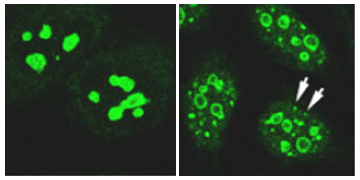
Cells expressing a fluorescent marker of the small ribosomal subunit. On the left, control cells showing the characteristic accumulation of these components in the nucleolus; on the right, RPS19-deficient cells showing the accumulation of aggregates in other regions of the nucleus, outside the nucleolus (arrows).
The use of high-content screening, a technology that acquires and analyzes thousands of images automatically using robotic microscopes, has been an important factor in the completion of this study. “Being able to use this technology allowed us to obtain a great deal of information not only about the effect of ‘switching off’ each of these genes on preribosome assembly, but also about the similarity or potential interdependence between them. We are excited to have this type of technology available at the IIBM in the near future,” says Miguel Sánchez.
The findings provide a broader view of how defects in the formation of the small ribosomal subunit alter the organization and balance of ribosomal components within the cell nucleus, and provide new insights into the molecular mechanisms that may contribute to the development of ribosomopathies.
Article reference: Gaspar, S. G., Sánchez-Álvarez, M., Ramírez-Cota, R. M., Nieto, B., Flujas, M., del Pozo, M. Á., Bustelo, X. R., & Dosil, M. (2026). Convergent nuclear proteostasis alterations across 40S ribosomal protein deficiencies. Nature Communications, 17, 8780. https://www.nature.com/articles/s41467-026-75682-6