Understanding how obesity affects the brain and whether these changes can be reversed is one of the major challenges in the development of new therapeutic strategies. A team from the Instituto de Investigaciones Biomédicas Sols-Morreale (IIBM), CSIC-UAM, has shown that an experimental treatment not only reduces body weight but also attenuates brain inflammation and partially reverses some of the alterations associated with obesity in a murine model.
The study, published in the journal Molecular Medicine under the title Acute anti-obesity treatment with celastrol reduces body weight, cerebral inflammation and metabolic imbalances in mice, was led by Blanca Lizarbe Serra, researcher of the Biomedical and Biophysical MR lab Group at the IIBM. Adriana Ferreiro, a predoctoral researcher at the Institute, is the first author of the paper. The study was also carried out in collaboration with the IIBM Preclinical NeuroImaging Group and the Stem Cells, Immunity and Cancer Group at the Universidad Complutense de Madrid.
The research focuses on celastrol, a natural compound with anti-inflammatory properties obtained from the plant Tripterygium wilfordii. To investigate its effects, the researchers used a murine model of diet-induced obesity and combined several techniques, including diffusion MRI, spectroscopy, immunofluorescence and hormonal analyses. This multimodal approach enabled a comprehensive characterization of the changes occurring in the brain following treatment.
The results show that the treatment significantly reduces body weight and attenuates obesity-associated brain inflammation, particularly in the hypothalamus. In addition, it partially reverses several microstructural and metabolic alterations detected in the animals' brains. Specifically, MRI-derived maps showed normalization of different parameters in this brain region, while microglia and astrocytes recovered a less reactive morphology, indicating a reduced inflammatory response.
Furthermore, spectroscopy revealed a normalization of the concentration of several brain metabolites, together with a decrease in markers associated with glial proliferation, metabolic stress and oxidative stress.
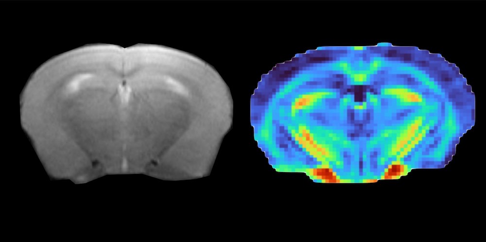
MRI image of a brain mouse treated with celastrol. The left panel shows the transverse relaxation time, while the right panel shows a fractional anisotropy parametric map used to evaluate changes in brain microstructure.
Taken together, these findings provide new evidence on the brain mechanisms involved in obesity and in the response to anti-obesity treatments. They also highlight the potential of MRI as a non-invasive tool to monitor brain inflammation in vivo and to assess the efficacy of future therapies targeting this disease.